
Indian Paediatrics & Critical Care
Loading QBank Portal & Clinical Guidelines...
Loading QBank Portal & Clinical Guidelines...
In summary:
Gross structure of the liver
Fine structure of the liver
Hepatic sinusoids
Anatomy of the hepatic circulation, briefly
Anatomy of the biliary circulation
ACUTE
LIEVR FAILURE è “fulminant
hepatic failure”
-
A
rare syndrome characterised
by rapid onset
of severe hepatic
dysfunction in the
absence of pre-existent
liver disease.
-
In ALF, massive or
submassive necrosis of
hepatocytes causes impairment
of hepatic synthetic
functions and development of hepatic
encephalopathy.
Acute Liver
Failure Study Group
(ALFSG) defines è
-
ALF
in adults as
acute hepatic dysfunction
(coagulopathy, poor hepatic synthetic function) with development of
encephalopathy within 8 weeks.
-
In children, encephalopathy is difficult
to assess, and may not
develop until terminal
stages.
-
Hence,
in children, ALF
as defined by
the Paediatric Acute
Liver Failure Study
Group (PALFSG) includes
–No history of known chronic
liver disease
–Evidence of
acute liver injury
(increased liver enzymes,
hyperbilirubinaemia),
i.e. evidence of liver dysfunction within 8 weeks of
onset of symptoms
–Coagulopathy unresponsive to
vitamin K
INR >1.5 in
patients with hepatic
encephalopathy, or
INR > 2.0 in patients
without encephalopathy).
Causes è
-
Aetiology
of ALF provides
one of the
best indicators of
prognosis, and also
dictates specific management options.
-
Severity
of coagulopathy and
possibility of recovery
also depends on the aetiology.
-
In developed nations drugs account for 70–80% of the causative factors,
whereas in developing
nations idiopathic and
viral infections account
for the majority
of the aetiological
factors.
Pathophysiology
è
-
injury
to hepatocytes causing cell
damage or cell
death by necrosis
or apoptosis (both may coexist
-
Acute Wilson disease and Reye syndrome àMitochondrial permeability
is increased and is associated
with apoptosis.
-
In
the early phase
of injury from
warm and cold
ischaemia, hepatocytes and
endothelial cells activate
the mitochondrial permeability
transition, and mitochondrial
injury resulting in ATP
depletion and ensuing necrosis.
-
Necrotic injury of liver cells leads to a loss
of cell membrane integrity and
eventual rupture and
cell death with
the release of
cytosolic proteins - LDH, ALT, AST and ferritin.
-
Cellular
glutathione is diminished, especially
following ALF with
acetaminophen. à
This increases cellular
susceptibility to oxidative
injury and impairs
the ability to
conjugate and detoxify some toxic substances.
-
Other detoxification mechanisms
are impaired, including the
transport systems for
bilirubin, leading to
cholestasis and conjugated hyperbilirubinaemia.
-
An important feature is the general loss of
Kupffer cell function that
results in reduced
clearance of endotoxin
and other substrates
regularly presented to
hepatocytes by means of the portal system, increasing the risk of infection.
-
Site
of injury within
the liver plays
an important role
in determining the
ability of the
liver to regenerate.
Injury to
the portal zone inhibits the
regenerative response, while
Injury to
the central zone
with sparing of
the portal zone permits more
frequent spontaneous recovery.
-
central
zone is more
susceptible to ischaemic injury.
Pathophysiological
Effects è
• Increased cardiac
output with decreased
systemic vascular resistance
• Increased portal pressure
• Renal vasoconstriction
• Pulmonary shunting; V/Q mismatch
• Total body fluid overload
-
LF
leads to the
development of severe
systemic inflammatory response
syndrome (SIRS) and extensive energy expenditure with catabolism.
-
Rapidly
progressive, severe multiorgan
failure with unpredictable
complications such as:
1.
Cerebral
oedema/intracranial hypertension —varying
grades of encephalopathy
2.
Infection—septic shock, peritonitis, etc.
3.
Coagulopathy —GI
bleeds, mucosal bleeds,
etc.
4.
Haemodynamics—vasodilatory shock, hypovolaemia
5.
Renal
failure—pre-renal failure, ATN,
hepatorenal syndrome
6.
Metabolic
concerns—hypoglycaemia,
dyselectrolytaemia
-
Most critical element to patient survival with
ALF is its
early recognition.
MANAGEMENT è
-
A multisystem disease with an unpredictable
course.
-
Mortality
in ALF is
predominantly due to
raised intracranial pressure
(ICP), infections and
multiorgan failure.
-
GOAL à Management of
these conditions while
awaiting recovery of
liver function or
liver transplantation (LT).
General
Measures à
-
Nursed in a quiet environment, preferably in PICU.
-
Put central
venous line à measure
central venous pressure,
administer fluids, medications
and blood products,
and collect blood samples.
-
Volume
resuscitation - aggressively if
required.
-
Iv
at three-fourth maintenance
requirement.
-
Fluids - glucose based with a
glucose infusion rate
of at least
6–8 mg/kg/min.
-
Vasoactive drugs
such as dopamine
and dobutamine, if clinically
indicated.
-
Strict intake and output charting should be
done.
-
NG tube - feeding/drainage
-
Urinary
catheter for output
measurement.
-
Care
of bowel, back,
bladder, skin and
eyes should be done.
-
Following
monitoring should be
done:
(i)
vital
signs, every 4
hours, more frequently
in an unstable
child;
(ii)
neurological
observations/coma grading, electrolyte,
arterial blood gases,
blood sugar and
prothrombin time every
12 hours (more
frequently in an
unstable child);
(iii)
daily
measurements of liver
span and prescription review;
(iv)
liver
function tests, blood
urea, serum creatinine,
calcium and phosphate
at least twice
weekly; and
(v)
Surveillance blood and urine cultures during the
course of illness.
Aetiology-Specific
Therapy è
Paracetamol
(Acetaminophen) Poisoning
-
Rummack–Mathew nomogram may aid
in determining the
likelihood of serious
liver damage, but
cannot be used
to exclude possible
toxicity due to
multiple doses over
time, when the time
of ingestion is
unknown.
-
Complete
recovery occurs with
timely (started within
24 hours) supportive
care and N-acetylcysteine (NAC).
-
NAC has
proven role in
the treatment of
paracetamol toxicity.
-
It
should be given
as early as
possible, but may still be of value 48 hours or more after ingestion.
-
Intravenous
administration (loading dose
150 mg/kg in
5% dextrose, followed by a continuous infusion at 10
mg/kg/h).
-
Continue
NAC till hepatic
function normalises (as evidenced by decreasing INR).
-
Oral
charcoal is given
if patient presents
within 2 hours
of ingestion. It
does not interfere
with absorption of oral NAC.
Non–Paracetamol-Induced
ALF
-
NAC
is useful in
non-paracetamol ALF also
(as it improves
systemic haemodynamics and
tissue oxygen delivery).
However, the probability of complete recovery is less than that with
paracetamol-induced hepatic injury.
-
Viral hepatitis A
(and E)–related ALF
must be treated
with supportive care
as no
virus-specific treatment has
been proven to
be effective.
-
Nucleos(t)ide
analogues should be
considered for hepatitis
B–associated ALF and
for prevention of
post-transplant recurrence.
-
Patients
with known or
suspected herpes virus
or varicella zoster
as the cause
of ALF should
be treated with acyclovir (5–10
mg/kg IV every
8 hours) and may be considered for LT.
-
Patients
in whom Wilson
disease is the
likely cause of
ALF must be
promptly considered for LT.
-
Patients
with autoimmune hepatitis
should be considered
for LT even
while corticosteroids are
being administered.
Intensive
Care Issues in Acute Liver Failure è
• Hepatic encephalopathy/cerebral oedema
• Infection
• Coagulopathy
• Haemodynamics
• Renal failure
• Metabolic concerns
Hepatic
Encephalopathy è
-
Most
serious and life-threatening complication
of ALF.
-
Occurrence
of cerebral oedema
and intracranial hypertension
is related to
severity of encephalopathy (risk
of cerebral oedema
25–35% in Grade
III HE; increases to 65–75% in Grade IV).
Pathophysiology of Hepatic Encephalopathy
-
Increased
ICP is a
major cause of
mortality in patients
who have ALF.
-
Hyperammonaemia, increased cerebral
blood flow and
enhanced inflammatory response play
central roles.
-
Hyperammonaemia is
associated with increased
levels of glutamine
within astrocytes.
-
Accumulation
of glutamine results
in cell swelling and brain oedema.
-
Hyperammonaemia alone does
not predict intracranial hypertension
-
Increased
cerebral blood flow
may contribute to
the development of
cerebral oedema by
facilitating water movement
down altered osmotic
gradients generated by
increased concentration of
glutamine within astrocytes.
-
In
addition, markers of
inflammation are increased
in ALF, perhaps
reflecting the SIRS.
SIRS seems to
result in worsening
encephalopathy, although its
direct relationship to brain swelling is not defined.
-
HE
encompasses many neuropsychiatric disturbances, ranging
from minor confusion
and disorientation to
frank coma, resulting
in intracranial hypertension.
Prevention/Management
of Cerebral Oedema in ALF
-
• Grade I/II encephalopathy
–Consider transfer to LT centre
–CT brain to rule out other causes of
decreased mental status–Avoid sedation/excessive stimulation
–Negative fluid
balance as tendency
to retain water
–Antibiotics: Surveillance
and treatment of
infection. Prophylactic antibiotics
possibly helpful if HE rapidly
worsening
–Lactulose: Possibly helpful
–No role for l-ornithine–l-arginine)
-
• Grade III/IV encephalopathy
–Neuroprotective care–Intubate and
mechanically ventilate
–Negative fluid
balance as tendency
to retain water
–Consider placement
of ICP monitor:
Target therapy to
maintain cerebral perfusion
pressure (CPP)
–Seizure control; role of prophylaxis
unclear
–Mannitol and
hyperventilation as rescue
measures only
–Therapeutic hypothermia: Promising
results
• ICP monitor
insertion: An ongoing debate
-
Benefit of ICP monitoring
-
Allows
early recognition of
intracranial hypertension
-
Helps
target therapy to
CPP (CPP =
MAP – ICP)
-
Most
useful in patients
with Grade III/IV
HE
-
Particularly
useful during orthotopic
LT when shifts
in haemodynamics can
cause large fluctuations in CPP
-
Target
CPP >45 mmHg
in infants, >50–55
mmHg for older kids and > 60 mmHg for adults.
-
Risks of ICP monitoring
-
Intracranial
bleed: 10–20% depending
on the type
of ICP probe
used. Correct coagulopathy
prior to insertion
till INR <1.5.
Recombinant factor VII
concentrate (rFVIIa) may
be helpful during
the procedure of ICP monitor
insertion
-
Infection
-
Correction
of coagulopathy should
be continued for about 48 hours
after insertion of ICP probe to prevent bleeding.
-
Measures
to decrease ammonia:
Serial trends of
serum ammonia level
important for risk
stratification.
-
Serum ammonia > 200 μmol/L are usually
associated with herniation
–Drugs used to decrease ammonia level
Non-absorbable disaccharides: Lactulose/lactilol
–Acts as
a laxative; decreases
intestinal absorption of ammonia
and glutamine
–Risk of gaseous bowel distension
–Can be given per rectally as enema
–Cannot be used in patient with ileus
–Insufficient evidence of effect
Antibiotics
–Reduces intestinal ammonia
production by acting against urease-producing microorganisms
–Rifaximin more
effective than neomycin/metronidazole. Less effective than
lactulose -Zinc
–Regulation of protein and nitrogen
metabolism
–Slow
acting; more useful
in chronic hepatic
failure
Steroids: No role
Therapeutic hypothermia
–Gaining acceptance
as a measure
to reduce refractory ICP in ALF
–Moderate hypothermia (32–34°C)
↓ cerebral metabolism
Suppresses seizures
Normalises cerebral blood flow
↓ delivery of ammonia to brain
↓ oxidative stress to brain
Reversal of SIRS
Infections in
ALF
-
• ALF
is an immunocompromised state
due to decreased Kupffer cell function.
Hence, patients are prone to
bacterial and fungal
infections.
-
Common
organisms include Staphylococcus, Streptococcus and GNB.
Broad-spectrum coverage with a third-generation
cephalosporin, vancomycin/teicoplanin
and fluconazole is
recommended wherever indicated.
-
• Peripheral
vasodilatation due to
ALF closely mimics septic shock.
-
• Since occult infection
is a common
cause of worsening encephalopathy, periodic
surveillance cultures to detect bacterial and fungal infections
as early
as possible should
be done, particularly
in patients who are likely transplant candidates.
-
Prophylactic
antibiotics and antifungals
are indicated only if the
patient has
–Hypotension
–SIRS with features of sepsis
–Signs of peritonitis
–Progression of encephalopathy
–Surveillance cultures
growing significant isolates
–Waiting for organ transplant.
Correction of
Coagulopathy in ALF è
-
• Coagulopathy
is a dynamic
marker of hepatic
dysfunction.
-
Risk
of bleeding increases
due to concomitant thrombocytopenia, hypofibrinogenaemia and
vitamin K deficiency.
-
Vitamin
K should be
prescribed for all
patients with ALF
and should be
given as an
infusion.
-
• Hallmark: Decreased synthesis of procoagulants
–Impaired anticoagulation and
fibrinolysis
–Thrombocytopenia and thrombasthenia
-
• Factor V has prognostic value.
-
• Indications of correction of coagulopathy
–Significant spontaneous bleed
–Prior to invasive procedures
–Profound coagulopathy (INR > 7)
Variceal
Bleeding and Haematamesis è
-
Major
gastrointestinal bleeding in
the setting of end-stage liver
disease and portal
hypertension may occur
because of oesophageal/gastric varices,
portal gastropathy or distal bowel varices.
Management à
-
• Resuscitate with colloid and blood products.
-
• Correct
coagulopathy (vitamin K,
FFP or cryoprecipitate).
-
• Start
IV octreotide or
any other octreotide
analogue that is available.
-
• Perform
gastroscopy ± colonoscopy with
a view to
confirming the cause
of bleeding and
endoscopic treatment (variceal
banding or glueing)
after the patient
has been resuscitated
and stabilised.
-
• If
bleeding persists and
oesophageal varices are
suspected or known,
consider inserting Sengstaken–Blakemore tube
if available, else use
the largest-sized Foley catheter into stomach and inflate balloon (as per
packet).
-
Apply gentle traction until there is resistance
(so that balloon abuts
gastro-oesophageal junction, compressing feeder vessels to oesophageal varices).
-
Take care not
to exert excessive
traction thereby pulling
balloon into oesophagus.
This may dislodge
clot or rupture
the oesophagus. Usually
this is rarely required and variceal banding and
glueing is undertaken
Ascites à
-
The
production of ascites
is complex and
related to portal hypertension.
-
Most children with chronic liver disease
and ALF will
present with ascites,
although it is rare as an acute presentation of ALF.
Management è
-
Thorough
clinical examination or
ultrasound if unsure.
-
Ultrasound
- distinguish ascites
from intraperitoneal blood
loss.
-
Rapid
development of increasing
ascites may indicate
spontaneous bacterial peritonitis (SBP).
-
Specific management is required only if
affecting feeding or ventilation
-
Use diuretics (spironolactone, frusemide).
-
• Maintain
albumin in the
normal range with infusion
of 20% albumin, giving a single dose of frusemide 1 mg/kg midway or
at end of infusion.
-
• If
there is respiratory compromise, consider peritoneal
tap and replace with IV
albumin 5 g/1000 mL of fluid removed.
-
Send a sample of the ascitic fluid for
microscopy, culture, cell count
and albumin for
surveillance of SBP.
Bacterial
Peritonitis è
-
Common
and severe complication of
cirrhotic patients with
ascites.
-
Common
organisms include E.
coli, Streptococcus viridans,
Staphylococcus aureus and
Enterococcus fecalis.
-
Clinical presentation - fever, leucocytosis, shock,
abdominal pain/tenderness or
only increasing ascites.
Management à
-
Peritoneal
tap for microscopy, culture
and cell counts.
-
Blood
cultures must also be obtained and sepsis workup should be
included.
-
Peritoneal
tap findings having
more than 250
polymorphonuclear cells/mL are
suggestive of infection
(calculate ratio for WBC to RBC if peritoneal tap is
blood stained).
-
• IV
antibiotics empirically must
be started.
-
Choice
usually is cefotaxime, and
changed based on cultures.
-
20% albumin IV may be needed at diagnosis
following tap.
-
Antibiotics
are continued for at least 5 days.
Haemodynamics
and Fluid Status in ALF è
-
• Hypovolaemia
and dehydration are important causes of precipitation of encephalopathy.
-
• Low
systemic venous return
(SVR) in ALF
leads to frequent
hypotension and bounding
peripheral pulses.
-
• If in shock, correct haemodynamics with one
or two isotonic fluid boluses, and start vasopressors/inotropes early.
-
Dopamine, noradrenaline and
adrenaline are useful.
-
• Avoid vasopressin in ALF; it
is associated with increase in
cerebral blood flow
and worsening I C P.
-
• Children
in ALF are more
prone for secondary
hyperaldosteronism
and so if
they are not
in shock then it
is preferable to
start the child
on two-third maintenance
fluid (isotonic dextrose
containing fluids).
-
Beware: Blood
products are a
common cause of
fluid overload.
-
Maintain
negative fluid balance yet euvolaemia.
-
Maintenance
of high normal serum
sodium (140–150 mEq/L)
helps in decreasing
cerebral oedema.
-
If
serum sodium is
persistently <140 mEq/L, restrict fluids further.
Renal
Insufficiency in ALF è
-
• Renal
failure is a
marker of poor
prognosis in ALF.
-
• Renal insufficiency in ALF can be due to
various reasons:
–Dehydration and pre-renal failure
–Acute tubular necrosis
–Hepatorenal syndrome
-
• Diuretics can precipitate hepatic encephalopathy by causing
hypovolaemia.
-
• Avoid nephrotoxic drugs and drug dosing has to be modified
accordingly.
-
• CRRT is the
best modality of
renal support in
ALF as it
helps to prevent
sudden haemodynamic changes
causing improved stability
of cardiovascular and
intracranial parameters.
-
Peritoneal dialysis
can be used
in smaller children and infants.
-
Common
metabolic derangements seen
in ALF include
hypoglycaemia, acidosis
or alkalosis, hypokalemia,
hypophosphataemia,
hypomagnesaemia, low ionised
calcium and poor
nutrition.
-
Nutritional
goals of a
patient with ALF:
(i)
calories,
100–120 kcal/kg/day; and
(ii)
Proteins,
1–1.5 g/kg/day. Role
of branched chain
amino acids (BCAA) is not clear.
-
• The
best way to
provide nutrition in ALF is
enterally.
-
If oral intake is not possible then only
TPN can
be started.
-
It
is preferable to
convert parenteral nutrition to
enteral as early as possible as there is a high risk of infections with TPN.
Liver Support
Systems è
-
–Plasmapheresis/double volume exchange
-
–Liver assist device: Non–cell based
(artificial)
Haemoperfusion
Molecular adsorbent
regenerating system (MARS)
-
–Liver assist device: Cell based (bioartificial)
HepatAssist: Porcine
ELAD: Human hepatoblastoma cells
Bioartificial liver
(BAL): Cryopreserved human hepatocytes
-
–Auxiliary LT
-
–Hepatocyte transplant
Liver
Transplantation è
-
Most
commonly used criteria
to predict poor outcome and need
for LT are King’s College Criteria.
-
survival
ranges from 70%
to 80%.
-
•
Listing of patient:
The decision to
list the patient
must be made
when the likelihood
of spontaneous recovery
is low and
the child has
not suffered irreversible
cerebral injury or
respiratory failure.
King’s
college criteria for liver transplantation è
-
Mitochondrial
disorders and haemophagocytic lymphohistiocytosis (HLH)
will usually not
benefit from LT.
-
Ischaemic
hepatitis usually has spontaneous recovery
and should not
be considered for LT.
-
Relative
and absolute contraindications to
transplantation include uncontrolled
infection or sepsis,
coma with irreversible
brain injury, active
or recent malignancy,
uncontrolled intracranial hypertension
or severe intracranial
haemorrhage and progressive
or end-stage extrahepatic disease or systemic disease
that will not be corrected
by LT. Inadequate
cardiac or pulmonary function of a transplant candidate
is a relative contraindication to major surgery.
-
• In view of the organ shortage, apart from
whole LT, split LT, grafts
from living donors
and extended criteria
donors are being
used
Miscellaneous
points è
-
Causes of a Raised ALT and AST
|
-
AST much higher than ALT -
Hepatic causes: -
Alcoholic hepatitis -
Alcohol toxicity -
Early ischaemic liver injury -
Extrahepatic causes: -
Rhabdomyolysis -
Myocardial infarction |
-
ALT much higher than AST -
Later in ischaemic liver injury -
Viral hepatitis -
Toxic hepatitis (eg. paracetamol) |
-
-
-
Nepean ICU
protocol for
handling this device.
1. Protect the airway.
Ideally, the patient should be intubated.
This prevents you from inserting the tube into the trachea accidentally, and
prevents aspiration of pooling oesophageal blood or displaced gastric content.
2. Inspect the tube and check the balloons for leaks.
LITFL also recommend to calculate the compliance of the balloon "by inflating the balloon with incremental 100ml
aliquots of air to maximal recommended volume (usually 250 -300ml for SBT,
450-500ml for Minnesota) and note the corresponding balloon pressure at
each step". This is
highly appealing to any person who enjoys graphs.
3. Lubricate the tube.
4. Position the patient sitting up to 45°
This protects them from aspiration
5. Insert the tube into the mouth or nose.
The college answer offers the nares as an option, but realistically everybody
always uses the orogastric route because these patients are always
coagulopathic and thrombocytopenic from their chronic liver disease. Moreover,
the tube is huge and thick, with big balloons- they will shred the nasal mucosa
on the way in regardless of how much lube you cake them in. The insertion
should ideally be performed under direct laryngoscopy so that you can be sure
you are in the oesophagus.
6. The tube should be advanced to 50cm.
The college answer prescribes a depth of 50cm, which is consistent with the
classical technique for insertion (Bauer et al, 1974). The alternative is to measure from
mouth to angle of the jaw, then suprasternal notch and xiphisternum. LITFL
authors recommend the latter method, acknowledging that humans vary in the
length of their oesophagus.
7. Inflate the gastric balloon. Check position with a
chest Xray.
There seems to be some disagreement as to how much one might inflate. The
college recommend 250ml; LITFL mention that the Minnesota tube should take
450-500ml. Locally, we are more cautions: we inflate with about 100ml and then
check position with an AXR. If one has produced a compliance curve for their
balloon, one may check the balloon pressure against their curve to see whether
it has been inflated in the oesophagus (LITFL offer a 15mmHg increase in
pressure as a rough guide: if the post-insertion pressure for a given volume is
more than 15mmHg higher than the pre-insertion pressure, then the balloon needs
to be repositioned as it is likely in the oesophagus. )
8. Withdraw the tube until resistance is felt (at 30-35cm)
This is usually the depth to the gastro-oesophageal junction. Tension develops,
which gives one the impression that the balloon is up against an obstacle of
some sort. If one has not inflated with enough air there will be no resistance,
and the balloon will come out of the mouth to theembarrassment of the
operator.
9. Aspirate the gastric and oesophageal ports.
If there was vigorous bleeding, it should have stopped by balloon tension.
10. Decide whether or not to inflate the oesophageal
balloon.
If you already know where the varices are on the basis of a gastroscopy result,
you may use your judgment (i.e. there is no point of inflating the oesophageal
balloon for gastric varices). Otherwise, one is guided by blood loss. If
bleeding from oesophageal and gastric ports has ceased, then you may
leave oesophageal balloon deflated. Bauer et al (1974) recommend to irrigate the suction ports
with warm saline, to assure oneself that the aspirate returns clear and that
there is no new bleeding.
11. If appropriate, inflate the oesophageal balloon to
25-30 mmHg pressure.
The maximum oesophageal pressure is 40mmHg. If the bleeding in the oesophagus
has stopped, one should deflate the oesophageal balloon by 10mmHg every 2
hours.
12. Apply traction to the tubing
The precise amount of traction is uncertain. Some centres specify 1kg, others
2kg. The college answer calls for a 500ml bag of fluid, suspended over a
pulley.
-
Link to join DTaP PICU Group è
https://t.me/+in4hkN3oWCUxMmY1
-
Link to join DTa Ppedia à https://t.me/+C17Q8ftUCrM2Y2Rl
STATION 10
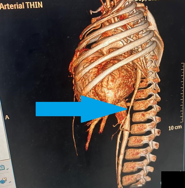
A previously healthy 6 year old
female came with C/O fever for 2wk, and difficulty in breathing for last 3
days, on admission pt had tachycardia, tachypnoea with feeble femoral pulses
and 2-D Echo show Dilated left ventricle with Ejection fraction
<15%, BP in Upper limb was 134/96 while in Lower limb 72/54. A
diagnostic imaging modality was done shown below in figure. Look at the figure
and answer the following?
Marks
Q 1)
What is this diagnostic imaging modality
?
1
Q 2)
What is the arrow showing in above
depicted picture? 1
Q 3)
Identify the diagnosis ?
1
Q 4)
Write any two differential diagnosis of
above condition ?
0.5*2=1
Q 5)
Write any one management option.
1
At a basic level this is a condition which results from the cerebral oedema that occurs when an ineffective osmole is enthusiastically extracted from the extracellular fluid by dialysis. In this, it resembles the cerebral oedema that can develop with the rapid overcorrection of glucose in a patient with a hyperosmolar hyperglycaemic state. Its one of those rare disease states that, when first recognised, was immediately and fully understood by the authors of the seminal article, with little else added in the subsequent years.
That seminal paper was Kennedy et al (1962), and is really just a letter to the editor of the Lancet. This group of clinicians noted that among renal failure patients with uraemia, a group failed to improve following dialysis - instead developing worsening confusion even while their biochemistry looked better. They made the connection between this phenomenon and the urea concentration, taking a hint from contemporary neurosurgical practices (where at the time it was routine to give doses of urea for cerebral oedema, in the same way as in the modern era one might give mannitol). Coming to the conclusion that some sort of a reverse version of this effect must have been operating in their patients, they followed this up with a series of experiments, demonstrating the lag between blood urea and CSF urea concentrations. In this manner, Kennedy et al were able to demonstrate that a concentration gradient is established during and after dialysis.

Cold Shock: Dopamine / Adrenaline first-line. Warm Shock: Noradrenaline first-line.
PALICC-2 Criteria: Oxygenation Index (OI) = (MAP x FiO2 x 100) / PaO2.